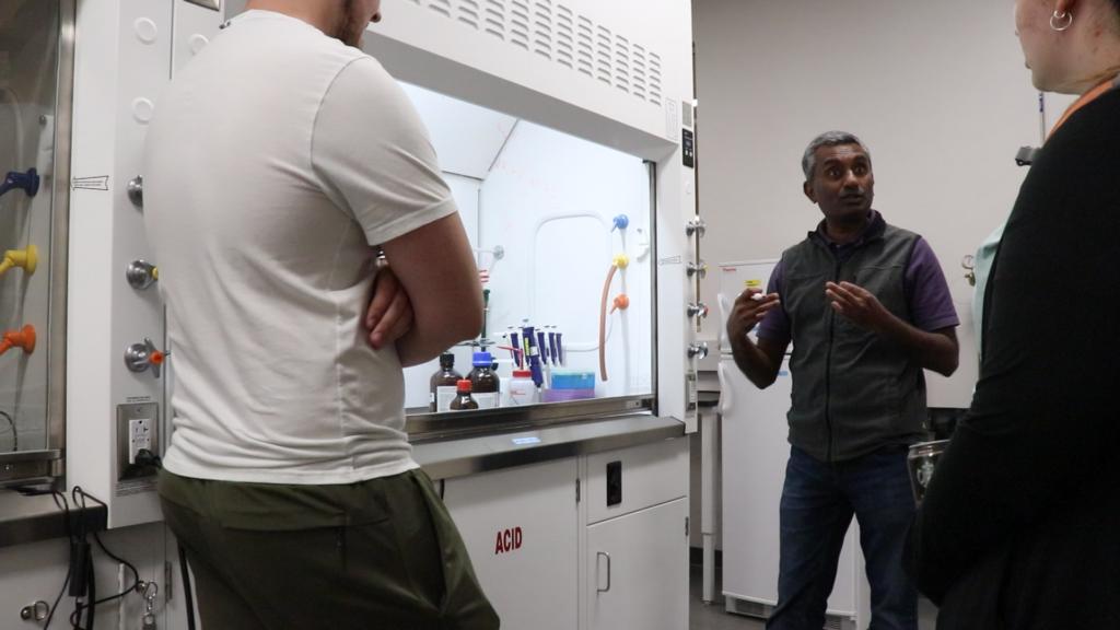
Apadaca’s 353 chemistry lab is home to lots of research equipment, chemical substances along with hardworking students and faculty.
Dr. Channa De Silva, chemistry professor, is the faculty advisor for one section of lab. The focus is on nanotechnology.
What is nanotechnology?
As De Silva explains, nanotechnology is not new but today its applications can bring new revolutionary discoveries in many areas and sciences.
“It is a type of technology that deals with science and engineering aspects of materials at the nanometer region,” De Silva explained.
This means that nanoparticles are extremely small, actually between 1 to 100 nanometers in size.
To put this into perspective, one nanometer equals a billionth of a meter.
De Silva believes that although they are small, they can make a big impact on the world, specifically the field of biotechnology which includes medicine, clinical and pharmaceuticals.
“Nanotechnology is important because you can manipulate the materials at the nanometer level,” said Dr. De Silva.
He explains that the particles small scale leads them to have multiple applications, as long as they can be manipulated.
De Silva began working with nanotechnology in 2006, during his doctoral studies at the University of Arizona.
He first became interested in the field because it was not yet well developed. It is still an emerging field in 2022 with lots of possibilities.
“We wanted to contribute to the science and make it accessible in the future,” De Silva said of his initial interest in the field.
De Silva is adamant about the potential of this technology, but he does stress that the toxicity of nanoparticles has not yet been tested. Before they can be put to the market, this will have to be tested.
De Silva says some scientists are working on their toxic effects, but he believes that more needs to be done. He explained that his students do study nanotechnologies toxic effects on a small scale, but he stressed that it needs to be expanded.
“We have recognized that importance and that is another direction we may move in the near future,” De Silva said.
What the team is working on
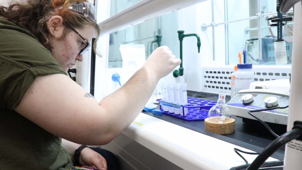
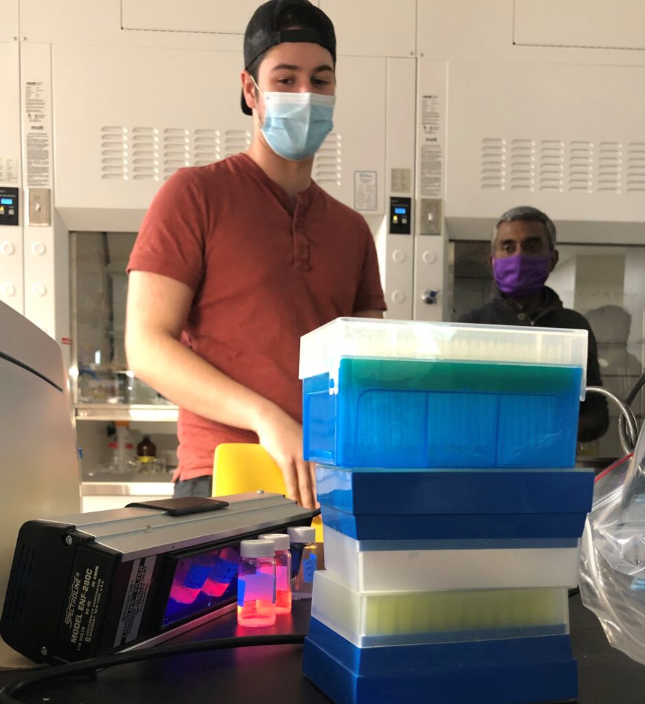
De Silva began working on the lab’s main project, the use of nanotechnology in cancer imaging, back in 2014. Currently, he has 10 students working on the project.
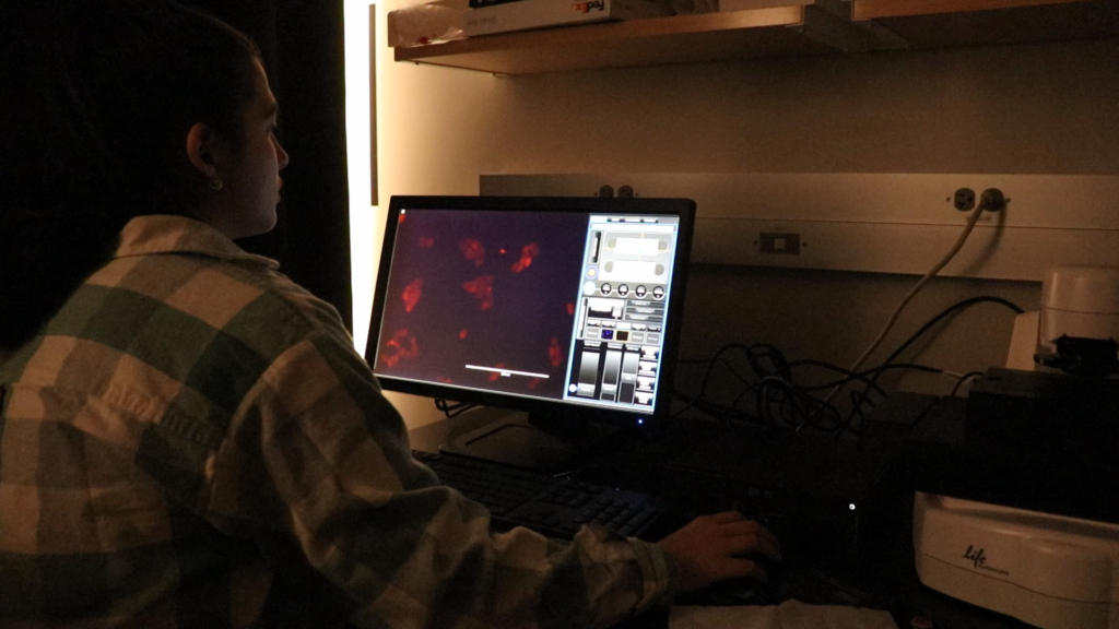
De Silva and his students believe their research could lead to high light-emitting nanoparticles used every day in the medical world detecting and identifying certain types of human cells responsible for certain diseases or conditions such as cancer.
He and his team of students are working with a few different types of nanoparticles that all serve different purposes. One of the combinations that the lab is working with is Calcium Fluoride and Europium.
When placed under UV light, Europium emits a monochromatic light or a pure red color.
One such application for this combination is cancer imaging.
For these nanoparticles to work the way De Silva and his students want, they must create a peptide.
Megan Rathbone and Yailyn Camelia are biology students working in De Silva’s Chemistry lab. Their project is to develop a peptide that De Silva’s students would use in their cancer imaging research.
Peptides are compounds of amino acids. As De Silva explains, the creation of a peptide is like making beaded necklaces. Each bead needs to go in the right order, or you would have made an entirely different necklace. The same goes with creating peptides.
The specific peptide that they are working on will be linked to melanoma, a type of skin cancer.
After this peptide ligand is created and is bound to the nanoparticles, cell experimentation will begin. A team of students will grow human cells and then inject nanoparticles within them to see if the peptide works.
In the video below, Dr. De Silva and Megan Rathbone describe how this peptide will work in their cancer imaging research and why it is important.
Another type of nanoparticle that the team is working on is zinc-oxide nanoparticles. These particles have anti-microbial properties which means they inhibit microorganism growth.
This type of technology would be very useful in hospital settings, military and field work where bacteria are teeming with life. The goal of this project is to one day create bandages that have zinc-oxide nanoparticles on them.
MacKenzie Gascon, a forensic chemisty major and rising senior, is also working in De Silva’s lab.
Her project is developing Terbium Calcium Fluoride nanoparticles that can be used for fingerprint imaging.
The hope is to place nanoparticles on finger print and shine a UV light on them, exposing the small details of the print that current technology cannot see.
Process of the cancer imaging project
The team’s cancer imaging project is very complex and has many moving parts.
The first step of the process took around 4 years. This part was called synthesis or developing methods of creating nanoparticles.
The video below details the step-by-step process of how Dr. De Silva’s students create these nanoparticles.
He explains that the process for creating these particles is like cooking.
The team worked to optimize the conditions of the experiment, such as the temperature of the scientific microwave, the time spent in the microwave, the power level of the microwave radiation, and the proportion of starting materials.
“It’s like coming up with a new recipe for a pizza that no one has ever made. It’s trial and error based. It’s something that no one has ever done before,” De Silva said.
Next the team moved to stage 2 of their process. Making the particles glow brighter.
To do this they modify the surface of the nanoparticle with other chemicals.
Now that the particles are finished the team moves to create the peptide. This is where the team is currently.
From there the team will couple the nanoparticles with the peptide at a molecular level.
Then the particles can be injected into human cells.
This part of the project De Silva must widen the scope as this kind of experimentation goes beyond the reaches of chemistry and into biology.
Yailyn Camelia is also working in Dr. Robert Youker’s biology lab, 429 in Apadaca, to grow and maintain the cells that will be used in the project when the team has reached that stage.
These nanoparticles could be a gamechanger not only for science but for how medicine is practiced in the future.
Dr. De Silva explains that while the world expects a lot from nanotechnology, it is still a new science, and still developing.
Currently, there are many researchers looking at different applications of nanotechnology. A few of those include paint, food packaging and fertilizer.
“It’s still in many research labs but the idea is to have products in the market in another 5 to 10 years,” said Dr. De Silva.
Even though this research is important to the world, Dr. De Silva never loses sight of what is in front of him.
“Our goal here is to teach our students the state-of-the-art synthetic methods and analytical methods so when they are graduating, they are ready for the future industry and higher education,” De Silva said.